- Supplier of advanced medical imaging systems
- 77500333 021
- 77504141 021

Phion 2.0

ImagePilot
فوریه 25, 2026
PACS Scalable Solution
فوریه 25, 2026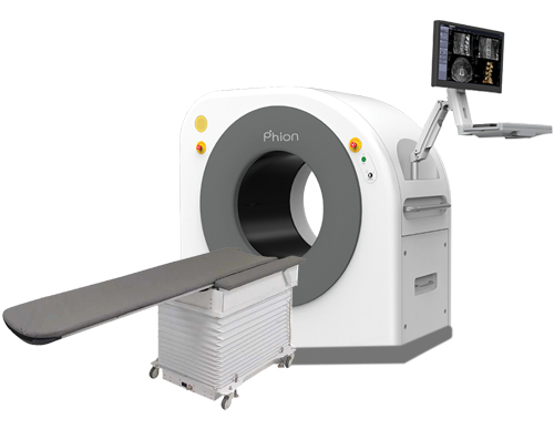
A NEW PIONEER OF CONE-BEAM CT
Brand-New Artificial Intelligent Technology Largest Bore and Field of View
What is Cone-Beam CT?
Cone beam computed tomography (CBCT) is a radiographic imaging technique that allows for accurate, three-dimensional imaging of hard tissue structures. CBCT is the most important method among the recently emerging medical diagnostic imaging techniques. Unlike conventional systems, these systems use cone beams instead of fan beams. Since these systems use a curved detector, they obtain a volumetric image of the patient instead of taking images slice by slice. CBCT has become increasingly important in treatment planning and diagnosis in orthopedics, neurosurgery, dentistry, otolaryngology, interventional radiology, and others

Phion CT
With the hardware and software tools provided by this technology, we will be faced with a very wide range of processed images. These images, which are taken with the help of cone beam and with only one radiation and in a time of about 7 seconds, have very wide advantages over digital radiography and multi-detector CT scans, the most important advantage of which is the time and radiation dose, which significantly reduces the absorbed dose of the patient and the staff of the imaging departments and is an important factor in the discussion of radiation protection. Another advantage of this device is its portability and small size compared to multi-detector CT scan devices and simple radiographs, which helps us to find a wide range of clinical and paraclinical applications in various hospital departments and mobile hospitals in wide areas of treatment and rehabilitation
Phion CT Features
Ability to scan various anatomical areas with a wide field of view • Designed for imaging the spine, skull, pelvis, shoulder, limbs and joints, jaw and face and teeth, paranasal sinuses, arthrography, interventional radiology and veterinary medicine.• Can be used in the operating room in neurosurgery, orthopedics and ENT procedures.
• High spatial resolution, which provides high quality images and high detection power in imaging hard tissue.
• Low radiation dose, about a quarter compared to multi-detector CT scans
• User-friendly software and variety in reconstruction algorithms by the Xelis 3D platform
• Imaging in about 7 seconds
• Reconstruction of axial, coronal, sagittal and 3D images by the software itself in 40 seconds
• Easy installation with city electricity and minimal radiation protection facilities

Dental CBCT
1- Performing specialized oral and dental CBCT with large FOV
2- Reducing positioning and exposure time compared to conventional devices
3- Providing images with isotropic pixels for radiological evaluations, measuring alveolar bone length, AVN, periodontal evaluations, etc.
4- Device with SUPINE positioning and orientation to reduce motion artifacts, suitable for elderly and physically limited patients.
5- Ease for the patient and physician for injection, cannulation of sialography, and performing interventional procedures.
